


Home / Albums / Natural History / Anatomy 50

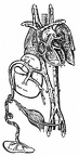 Plan of the foetal circulation
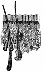 A cross section of the skin
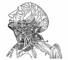 Lymphatics of the head and neck. B, the thoracic duct
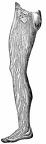 Lymphatics of the leg.
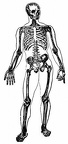 Skeleton
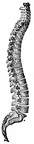 The Spine
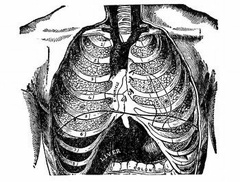 Front view of the thorax
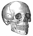 The Skull
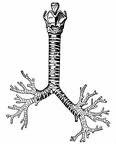 The cartilages of the larynx; the trachea and bronchi
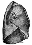 The root of the left lung
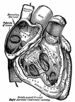 The right auricle and ventricle laid open
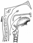 Passage into trachea and esophagus; Pharynx
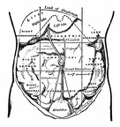 The regions of the abdomen and their contents
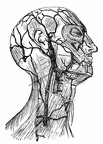 Superficial veins of the head and neck
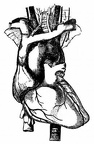 The arch of the aorta and its branches
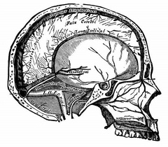 Vertical section of the skull, showing the sinuses of the dura mater
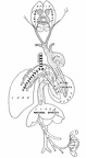 Illustrating Galen’s physiological teaching
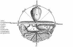 The Microcosm
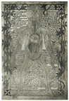 An anatomical diagram of about 1298
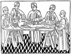 The first printed picture of dissection
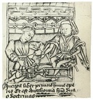 The figure shows a professor and pupil. The former is demonstrating the bones of a skeleton.
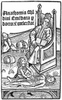 Title-page of Mellerstadt’s edition of the Anatomy of Mondino, Leipzig, 1493. The scene is laid in the open air
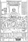 A dissection scene
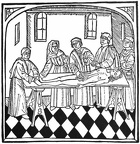 The first picture of dissection in an English-printed book
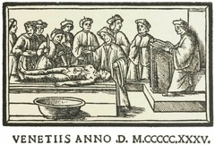 a lecture on anatomy
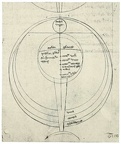 Roger Bacons diagram of the Eye
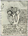 Leonardo Da Vincis diagram of the heart
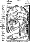 The figure shows the ten layers of the head
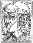 The layers of the head
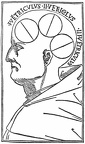 Venice, 1496, showing the ventricles of the brain
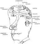 Diagram of the senses, the humours, the cerebral ventricles, and the intellectual facultie
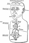 Illustrating the general ideas on anatomy current at the Renaissance
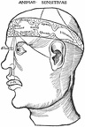 Diagram of the ventricles and the senses
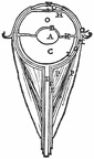 The Anatomy of the Eye
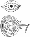 The Anatomy of the Eye
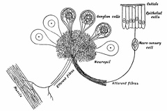 A Ganglion of a Leech
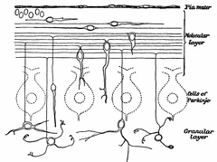 The Growth and Migration of Granules of the Cerebellum
 The Body of a Motor Neurone
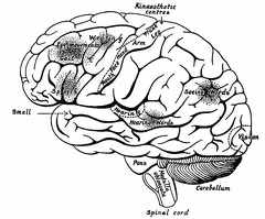 The Surface of the Left Cerebral Hemisphere, Cerebellum,and Medulla Oblongata.
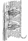 Highly Magnified Section through the Wall of a Circumvallate Papilla of the Tongue, showing Two Taste-Bulbs.
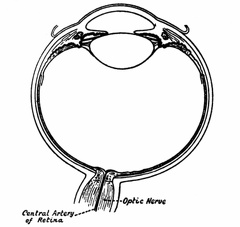 Horizontal Section through the Right Eye
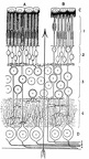 The Retina in Vertical Section
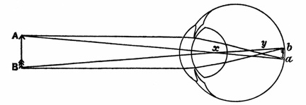 The Formation of an Image by the Refracting Media of the Eye
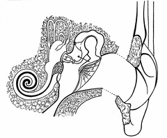 The External, Middle, and Internal Ear of the Left Side
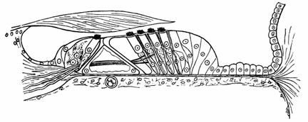 Organ of Corti
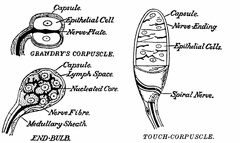 Sense-Organs susceptible to Pressure
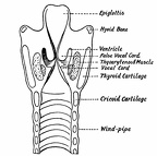 The Anterior Half of the Larynx seen from Behind
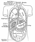 Diagram showing the Relative Positions of the Organs of the Chest and Abdomen.
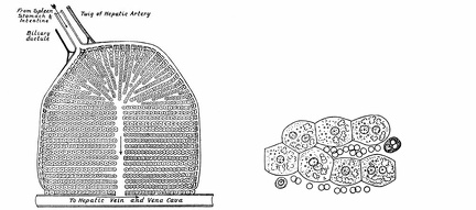 Diagram of a Lobule of the Liver
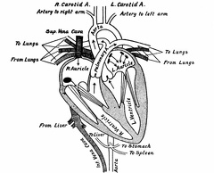 The Heart cut in the Plane of its Long Axis, and the Vessels which open into and out of it