


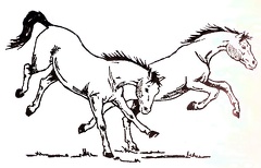
 Frolicking Horses
 The Horse
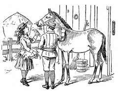 Feeding the horse
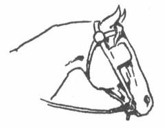 Comfort
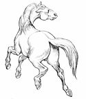 horse
horse Man riding horse
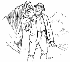 Man leading a horse
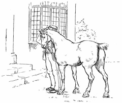 Man and horse outside a house
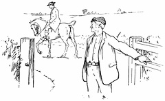 Going through the gate
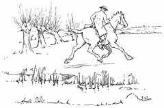 Trotting across a field
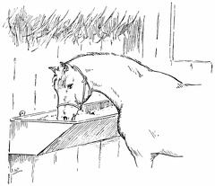 Feeding time
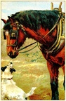 Horse and Dog
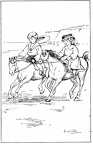 Two children riding ponies on the beach
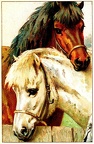 Two horses
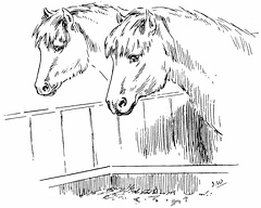 Two horses looking at their food
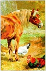 Horse and chickens
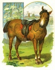 A horse
 Boy and Girl feeding a horse
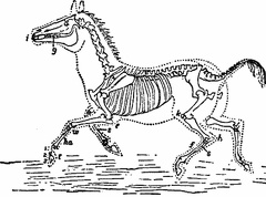 Skeleton of Horse
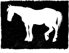 Horse
Horse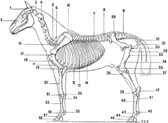 Skeleton of the Horse
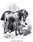 A new method of carrying dogs
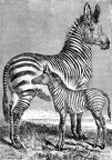 Zebra with young
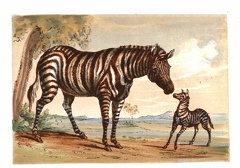 Zebra with young
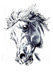 Terrified Horse
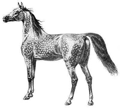 Speckled horse
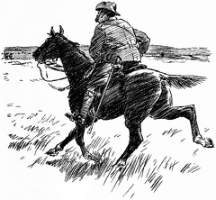 Soldier on horse
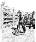 Rodeo Rider
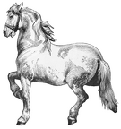 Prancing Horse
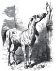 Need real food
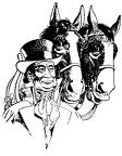 Man with two horses
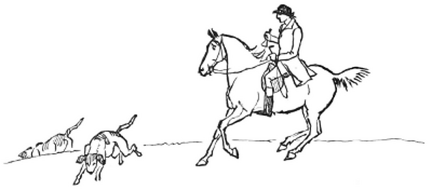 Hunting with the dogs
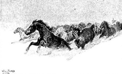 Horses running in snow
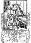 Horses in stall
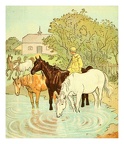 Horses Drinking
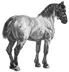 Horse
Horse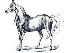 Horse
Horse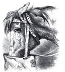 Horse with feedbag
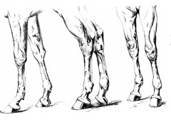 Horse legs
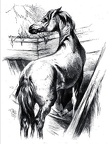 Horse in stall
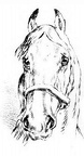 Horse Head
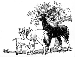 Horse family
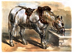 Horse drinking
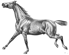 Horse cantering
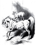 Horse and sheep show
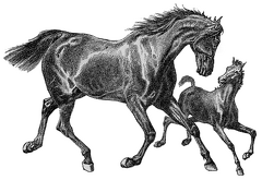 Horse and Foal
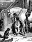 Horse and dogs ready for a ride
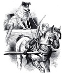 Horse and cart with dog driver
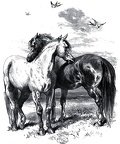 Horse affection
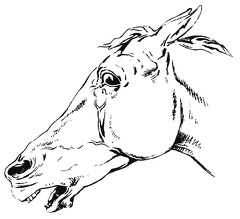 Frightened Horse
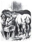 Feeding Time
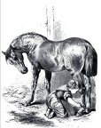 Child looking after horse
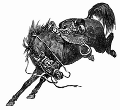 Bucking Horse
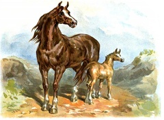 Brown horse and foal
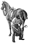 Blacksmith shoeing horse
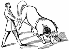 Bringing the horse to his knees
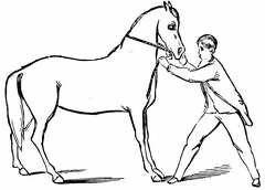 Teaching the horse to back
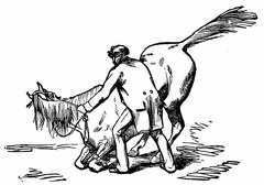 Preparing to lie down
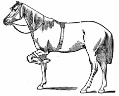 The short strap in use
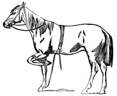 The short and the long straps