


Home / Albums / Natural History / Miscellaneous 19

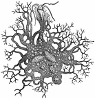 Protomyxa Feeding
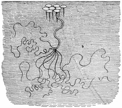 Fresh-Water Hydra
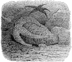 Star-fish Opening an Oyster
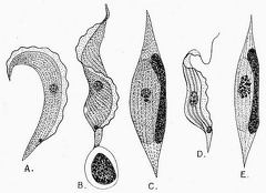 Trypanosoma Ziemanni, from the blood of the little owl
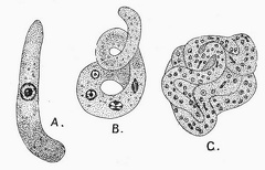 Trypanosoma Ziemanni, from the gut of the gnat
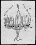 The Freshwater Jelly-fish of Regent’s Park (Limnocodium Sowerbii)
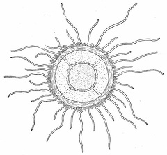 The Freshwater Jelly-fish of Lake Tanganyika
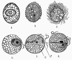 The unicellular parasite Benedenia, from the gut of the common Poulp or Octopus
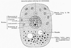 Diagrammatic representation of the structures present in a typical cell
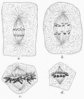 The Number of the Chromosomes
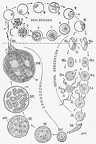 A diagram showing the life-history and migration of the Malaria parasite
 Lankesterella ranarum (Lank.), the parasite of the red blood-corpuscles of the edible Frog
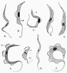 Various species of Trypanosoma from the blood of mammals, birds, and reptiles
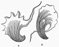 The earliest discovered Trypanosome, described by Gruby in 1843
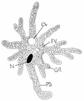 Diagram of amœba
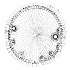 Volvox
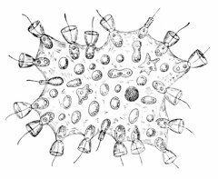 Proterospongia
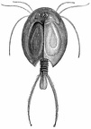 Gill foot
 Water flea