


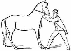
 Teaching the horse to back
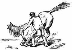 Preparing to lie down
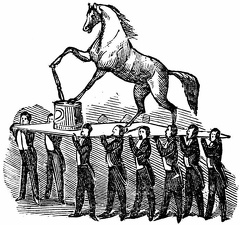 Pedestal Trick
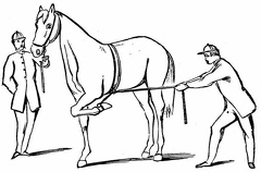 'Whoa'
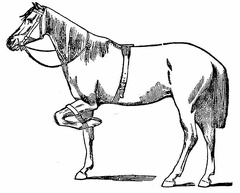 The short strap in use
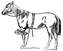 The short and the long straps
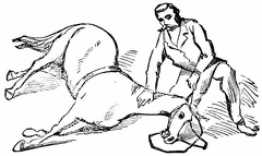 The Horse lying down
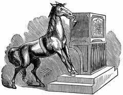 The Hand-organ performance
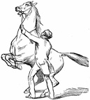 The application of both straps
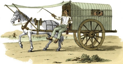 A Chinese Carriage
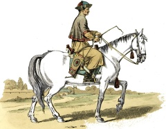 A Tartar Dragoon
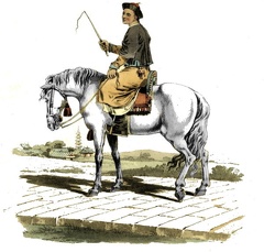 A mandarin's servant on horseback
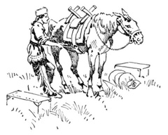 A Pack Horse
 Washington's Coach
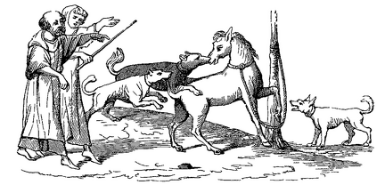 Fight between a horse and dogs
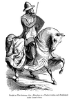 Knight in War Harness
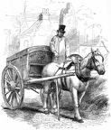 The Rubbish Carter
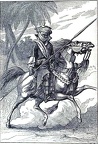 Member of the body-guard of the Sheikh of Bornou
 Lancer of the army of the Sultan of Begharmi
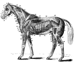 Muscles of the Horse
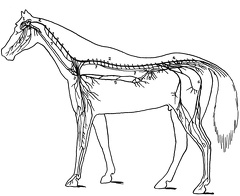 Nervous system of a horse
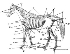 Skeleton of Horse
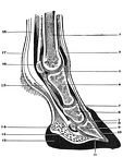 Cross section of foot of a horse
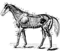 Deep muscles of the horse
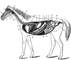 Digestive and Urinary apparatus
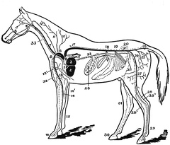 Heart and chief blod vessels