2 2020 »

 Portrait of Jean Cocteau
From an unpublished crayon sketch by Léon Bakst") Jean Cocteau
Jean Cocteau
Portrait of Jean Cocteau From an unpublished crayon sketch by Léon Bakst Young Lady Writing") Young Lady Writing
Young Lady Writing
Young Lady Writing A woman’s head
From the original drawing by Edwin Howland Blashfield") A Woman's Head
A Woman's Head
A woman’s head From the original drawing by Edwin Howland Blashfield three children looking at an aquarium
Any transparent vessel capable of holding water, even a Mas...") A self-sustaining or balanced aquarium
A self-sustaining or balanced aquarium
three children looking at an aquarium Any transparent vessel capable of holding water, even a Mason jar will make an aquarium from which a great deal of pleasure may be derived. The old way of maintaining aquaria in good condition required a great deal of care and attention. The water had to be changed at least once a day if running water was not available, and altogether they were so much trouble that as a rule owners soon tired of them. Modern aquaria are totally different. By a proper combination of fish and growing plants we can almost duplicate the conditions of nature and strike a balance so that the water need never be changed except when it becomes foul or to clean the glass. The wrong way to mount a horse—facing forward") The wrong way to mount a horse—facing forward
The wrong way to mount a horse—facing forward
The wrong way to mount a horse—facing forward Boy lying in a sleeping bag in the rain, without a tent.") With a head shelter and a sleeping bag he can keep dry and warm
With a head shelter and a sleeping bag he can keep dry and warm
Boy lying in a sleeping bag in the rain, without a tent. A heavy net is useful to capture aquarium specimens") A heavy net is useful to capture aquarium specimens
A heavy net is useful to capture aquarium specimens
A heavy net is useful to capture aquarium specimens A landing net should be a part of every fishing outfit. More fish are lost just as they are about to...") A landing net should be a part of every fisherman's outfit
A landing net should be a part of every fisherman's outfit
A landing net should be a part of every fishing outfit. More fish are lost just as they are about to be lifted from the water than at any other time. A gaff is used for this same purpose with fish too large to go into a landing net. A gaff is a large hook without a barb fastened into a short pole. If you have no net or gaff and have succeeded in bringing a large fish up alongside the boat, try to reach under him and get a firm grip in his gills before you lift him on board. If it is a pickerel, look out for his needle-like teeth. Addressing the golf ball before starting the swing") Addressing
Addressing
Addressing the golf ball before starting the swing The simplest way to catch minnows is with a drop net. Take an iron ring or hoop such as children use...") An excellent device for catching minnows
An excellent device for catching minnows
The simplest way to catch minnows is with a drop net. Take an iron ring or hoop such as children use and sew to it a bag of cotton mosquito netting, half as deep as the diameter of the ring. Sew a weight in the bottom of the net to make it sink readily and fasten it to a pole. When we reach the place which the minnows frequent, such as the cove of a lake, we must proceed very cautiously, lowering the net into the water and then baiting it with bits of bread or meat, a very little at a time, until we see a school of bait darting here and there over the net. We must then give a quick lift without any hesitation and try to catch as many as possible from escaping over the sides. The minnow bucket should be close at hand to transfer them to and care must be used not to injure them or allow them to scale themselves in their efforts to escape. At the top of the swing") At the top of the swing
At the top of the swing
At the top of the swing Jumping fences is the highest art of horsemanship") Jumping fences is the highest art of horsemanship
Jumping fences is the highest art of horsemanship
Jumping fences is the highest art of horsemanship Just before the ball is struck") Just before the ball is struck
Just before the ball is struck
Just before the ball is struck The hockey player's costume") The hockey player's costume
The hockey player's costume
The hockey player's costume The position of the men on a team is generally as the diagram shows but for various plays other form...") The lineup
The lineup
The position of the men on a team is generally as the diagram shows but for various plays other formations are used, provided that they do not violate the rules, which specify just how many men must be in the lineup and how many are permitted behind the line. In mounting, stand on the left side and place the left foot in the stirrup. Swing the right leg ove...") The right way to mount—facing toward his tail
The right way to mount—facing toward his tail
In mounting, stand on the left side and place the left foot in the stirrup. Swing the right leg over the horse and find the right stirrup with the toe just as quickly as possible. Do not jerk a restless horse or otherwise betray your excitement if he starts. Let him see by your calmness that he too should be calm. Forest travellers are always on the lookout for peculiar landmarks that they will recognize if they ...") The Wilderness Traveller
The Wilderness Traveller
Forest travellers are always on the lookout for peculiar landmarks that they will recognize if they see them again. Oddly shaped trees, rocks, or stumps, the direction of watercourses and trails, the position of the sun, all these things will help us to find our way out of the woods when a less observing traveller who simply tries to remember the direction he has travelled may become terrified. Chordæ tendineæ attach the margins of the auriculo-ventricular valves to musculi papillares which ...") The Heart cut in the Plane of its Long Axis, and the Vessels which open into and out of it
The Heart cut in the Plane of its Long Axis, and the Vessels which open into and out of it
Chordæ tendineæ attach the margins of the auriculo-ventricular valves to musculi papillares which project from the inner aspect of each ventricle. Diagram of a Lobule of the Liver divided vertically through its Axis.
In its centre is a space,...") Diagram of a Lobule of the Liver
Diagram of a Lobule of the Liver
Diagram of a Lobule of the Liver divided vertically through its Axis. In its centre is a space, the intralobular vein, through which the blood falls into a branch of the hepatic vein, on its way to the heart. An interlobular branch of the portal vein, which brings the blood from the digestive organs, pours it by many smaller vessels over the surface of the lobule. It filters into the lobule through innumerable pseudo-capillary vessels, or spaces, between the radiating columns of liver-cells. Arterial blood is brought to the lobule by a twig of the hepatic artery. Bile is drained away from it by an affluent of the hepatic duct. In the lower part of the diagram seven liver-cells are shown, forming a divided column, magnified about 300 diameters. The cells are loaded with glycogen, and contain minute globules of fat. Red blood-corpuscles and two leucocytes are seen between the columns of liver-cells. One of the leucocytes has ingested two blood-corpuscles. The ribs from the first to the tenth have been cut across in the lateral line. The eleventh and twel...") Diagram showing the Relative Positions of the Organs of the Chest and Abdomen.
Diagram showing the Relative Positions of the Organs of the Chest and Abdomen.
The ribs from the first to the tenth have been cut across in the lateral line. The eleventh and twelfth ribs do not reach sufficiently far forwards to be cut. With the exception of a short segment near its junction with the ascending colon, the small intestine has been removed. The trachea is seen to divide into bronchi beneath the arch of the aorta. The right lung has three, the left two lobes. The kidneys are situated behind all the other viscera. On their upper ends rest the two suprarenal capsules. The lower edge of the right lobe of the liver follows closely the line of the ribs and costal cartilages. Below the left lobe of the liver the stomach comes to the anterior abdominal wall. The transverse colon (large intestine) comes to the anterior wall below the stomach. Below the latter the wall is in contact chiefly with coils of small intestine. The vermiform appendix rests on the posterior wall. Spleen and pancreas are not shown in the diagram. The drawing shows the folds of mucous membrane, the vocal cords, which stretch from the tips of the ...") The Anterior Half of the Larynx seen from Behind
The Anterior Half of the Larynx seen from Behind
The drawing shows the folds of mucous membrane, the vocal cords, which stretch from the tips of the arytenoid cartilages to the recess behind the median portion of the thyroid cartilage. To the outer side of each vocal cord is seen the thyro-arytenoid muscle (cut across), consisting of a broad outer portion, chiefly concerned in closing the glottis during the act of swallowing, and a smaller internal portion, which regulates the length and the thickness of the segment of the cord allowed to vibrate. All are formed on essentially the same plan; a fibrous capsule invests a group of epithelial cells a...") Sense-Organs susceptible to Pressure
Sense-Organs susceptible to Pressure
All are formed on essentially the same plan; a fibrous capsule invests a group of epithelial cells amongst which a nerve ramifies. The simplest form is known as a Grandry’s corpuscle-a nerve ending in one or two plates between two or three epithelial cells. These organs are found in great numbers in the bills of aquatic birds. If a duck is watched whilst it is gobbling mud at the margin of a pond, it will be seen to have a remarkable capacity for discriminating between the shells of small snails, which it can crush, and stones, which it needs to drop from its bill. Its bill is also provided with small Pacinian corpuscles. The spiral lamina, on the left of the drawing, gives attachment to the membrane of Corti, which stre...") Organ of Corti
Organ of Corti
The spiral lamina, on the left of the drawing, gives attachment to the membrane of Corti, which stretches to the opposite wall. Below the membrane is a bloodvessel which runs its whole length beneath the tunnel of Corti. The tunnel is formed by pillars—the inner on the left, the outer on the right—which meet above it. On the left of the inner pillar is a hair-cell; to the left of this a nerve-cell with two nuclei. To the right of the outer pillar is a space; to the right of this four hair-cells alternating with four supporting cells, which hold up the reticulated membrane through apertures in which the tufts of hairs project. Three nerve-fibres are seen in the spiral lamina; they cross the tunnel to ramify between the rows of outer hair-cells. The lamina tectoria rests upon the tufts of hairs. From right to left, the figure shows the concha and lobule of the ear in profile; the external meatu...") The External, Middle, and Internal Ear of the Left Side
The External, Middle, and Internal Ear of the Left Side
From right to left, the figure shows the concha and lobule of the ear in profile; the external meatus (abbreviated); the drum, divided vertically, its posterior half visible; the hammer-bone, with the tip of its long arm attached to the drum, an arrow indicating the point of attachment and line of action of the tensor tympani muscle; the anvil attached by a ligament to the bony wall of the middle ear; the stirrup, with its foot-plate almost filling the oval window; the labyrinth, with the three semicircular canals above, and the scala vestibuli below. The curled black line shows the situation of the scala media, or ductus cochleæ (which contains the organ of Corti). Pulsations of sound which move the membrana tympani are transmitted by the three bones to the oval window. They shake the perilymph, producing waves which travel along the scala vestibuli to the apex of the cochlea, whence they return by the scala tympani to the round window (if they do not take a shorter course through the ductus cochleæ). The Eustachian tube opens out of the lower part of the middle ear. x, The common centre of curvature (nodal point of the several media). Rays which pass through this p...") The Formation of an Image by the Refracting Media of the Eye
The Formation of an Image by the Refracting Media of the Eye
x, The common centre of curvature (nodal point of the several media). Rays which pass through this point are not deflected. y, The principal focus of the system. All rays which are parallel to the optic axis converge to this point. The image of the point A is formed at a, the spot at which a ray parallel with the optic axis meets an unbent ray—the image of B at b. A, after Exposure to Bright Light;
B, After Resting in the Dark.
The arrow shows the direction i...") The Retina in Vertical Section
The Retina in Vertical Section
A, after Exposure to Bright Light; B, After Resting in the Dark. The arrow shows the direction in which light traverses the retina. C, Retinal epithelium, with its pigmented fringe. 1, Layer of rods and cones, separated by the external limiting membrane from 2, the layer of the nuclei of the rods and cones. 3, The ganglion-cells of the retina, which are homologous with the cells of the afferent root of a spinal nerve. Their peripheral axons ramify beneath the sensory epithelium (rods and cones and their nucleus-bearing segments), their central axons in 4, the inner molecular layer. D, Collecting cells on the front of the retina; a a a, their axons which conduct impulses to the brain; b, an efferent fibre from the brain. The slight depression in the retina in the axis of the globe is the fovea centralis, or yellow spot;...") Horizontal Section through the Right Eye
Horizontal Section through the Right Eye
The slight depression in the retina in the axis of the globe is the fovea centralis, or yellow spot; the optic nerve pierces the ball to its inner or nasal side. The lens, with its suspensory ligament, separates the aqueous from the vitreous humour. On the front of the lens rests the iris, covered on its posterior surface with black pigment. On either side of the lens is seen a ciliary process, with the circular fibres of the ciliary muscle cut transversely, and its radiating fibres disposed as a fan. These sense-organs are groups of elongated epithelial cells, set vertically to the surface. Their ce...") Highly Magnified Section through the Wall of a Circumvallate Papilla of the Tongue, showing Two Taste-Bulbs.
Highly Magnified Section through the Wall of a Circumvallate Papilla of the Tongue, showing Two Taste-Bulbs.
These sense-organs are groups of elongated epithelial cells, set vertically to the surface. Their cells are of two kinds—the one fusiform, slender, bearing each a bristle-like process which projects through a minute pore left between the superficial cells of the general epithelium; the other thicker and wedge-shaped. Nerve-fibres are connected with the fusiform cells. Sensory areas are enclosed by broken lines; certain centres in the association-zones are marked by d...") The Surface of the Left Cerebral Hemisphere, Cerebellum,and Medulla Oblongata.
The Surface of the Left Cerebral Hemisphere, Cerebellum,and Medulla Oblongata.
Sensory areas are enclosed by broken lines; certain centres in the association-zones are marked by dots. The sensory area of smell is on the inner aspect of the brain; so also is the area of vision which borders the calcarine and retrocalcarine fissures, and only rarely extends on to the external surface, as shown in the diagram. The sensory area of hearing is largely hidden within the fossa of Sylvius, the opening into which is indicated by the dark line above it. The kinæsthetic-sensory areas for the various muscles of the body occupy the territory between the dotted line in front and the bottom of the fissure of Rolando behind. They do not extend on to the posterior wall of this fissure. It is impossible at present to define the boundaries of any of the centres in the association-zones. In its centre is a large clear spherical nucleus, with a nucleolus. The body-substance is prolonged ...") The Body of a Motor Neurone
The Body of a Motor Neurone
In its centre is a large clear spherical nucleus, with a nucleolus. The body-substance is prolonged into five dendrites and an axon. Neuro-fibrillæ are seen in dendrites and axon. They traverse the body of the cell in all directions, in little bundles which are separated by angular granules of stainable substance (tigroids). Half a dozen nuclei of as yet undeveloped granules are seen lying beneath the pia mater. From this l...") The Growth and Migration of Granules of the Cerebellum
The Growth and Migration of Granules of the Cerebellum
Half a dozen nuclei of as yet undeveloped granules are seen lying beneath the pia mater. From this level to the bottom of the drawing granules are shown in successive stages of growth. These developing granules, selected from various preparations of the cortex of the cerebellum, were drawn from nature. Pear-shaped cells are set round a felt-work of nerve-fibrils (neuropil). A neuro-sensory cell is sho...") A Ganglion of a Leech
A Ganglion of a Leech
Pear-shaped cells are set round a felt-work of nerve-fibrils (neuropil). A neuro-sensory cell is shown with one fibre directed peripherally, branching on the surface; and one directed centrally, ramifying in the neuropil. Several very slender fibrils from the neuropil pass up the stalk of each ganglion-cell. They join a network near its surface. This net is connected by radiating fibrils with a coarser net which surrounds the nucleus. From the central net a relatively stout fibril passes to muscle-fibres. Note the diagonal line in the small diagram of the figure below. It is actually straight, but the ve...") Optical Illusion in dress
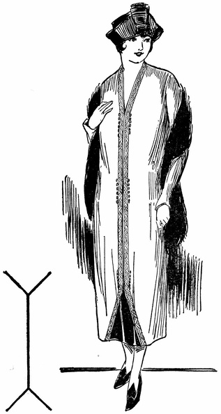
Optical Illusion in dress
Note the diagonal line in the small diagram of the figure below. It is actually straight, but the vertical lines which break it give it a “going-down-steps” appearance. This principle is used in the dress below—the two vertical panels of trimming break the line of the tunic and give the whole figure a more slender appearance than in the figure above.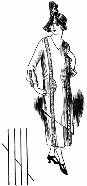
 Here a small all-over pattern minimizes size, the plaits and tassels lengthen, the necklace adds a s...") Choice of fabric
Choice of fabric
Here a small all-over pattern minimizes size, the plaits and tassels lengthen, the necklace adds a slenderizing touch. The appearance as a whole is graceful and youthful. Here trimming is used on two entirely different types of hats to give in each case added height to t...") Hats 2
Hats 2
Here trimming is used on two entirely different types of hats to give in each case added height to the figure and help in attaining a slenderizing appearance. Left—Hats with medium brims and high trimming are often becoming, especially if wide enough to avoid the pyramid effect. Right—High built trimming and delicate veils are advantageous where a double chin is the handicap. These two examples show how even a hat with drooping brim, if not too wide, can be worn by the stout...") Hats 1
Hats 1
These two examples show how even a hat with drooping brim, if not too wide, can be worn by the stout person if trimming is adeptly used to direct the vision upward and lend an illusion of height. Would you believe that the pattern of these two dresses is exactly the same? This illustrates how yo...") Two looks - same pattern
Two looks - same pattern
Would you believe that the pattern of these two dresses is exactly the same? This illustrates how you can vary a dress once you find the foundation lines that are becoming to you. One pattern can suffice for both a tailored and an afternoon dress, as you see both effects are pleasing in their slenderness. These two pictures illustrate improper and proper choice of fabrics for a stout figure. Above, the l...") Choice of fabric 1
Choice of fabric 1
These two pictures illustrate improper and proper choice of fabrics for a stout figure. Above, the large-figured material adds size, the fur trim shortens, the round beads shorten the neck. All conspire to emphasize weight.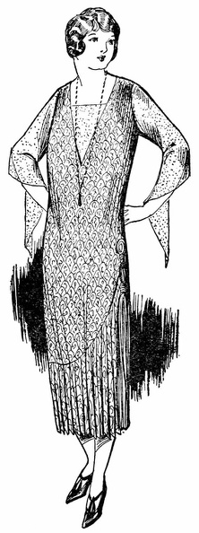
 Hats and shoes in these two pictures also illustrate incorrect and correct choice. The wide hat and ...") Plaits 2
Plaits 2
Hats and shoes in these two pictures also illustrate incorrect and correct choice. The wide hat and prominent straps below emphasize width and weight; the neat hat and cross-strap slippers here help to slenderize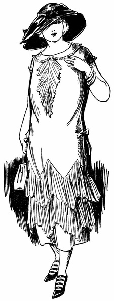
 When styles call for plaits, plaits may be used, but not in widening flares as shown here, rather in...") Plaits 1
Plaits 1
When styles call for plaits, plaits may be used, but not in widening flares as shown here, rather in slenderizing length lines as shown below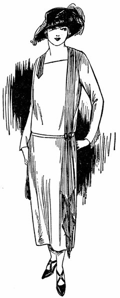
 The oblique line in the figure is made to seem longer and more graceful than the dress below by the...") Optical Illusion in dress
Optical Illusion in dress
The oblique line in the figure is made to seem longer and more graceful than the dress below by the parallel vertical lines of embroidery which intersect it and so emphasize its appearance of length and grace.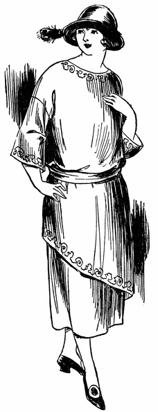
 Now see how the woman in the other picture has unknowingly emphasized her stoutness while the one in...") Optical Illusion in dress
Optical Illusion in dress
Now see how the woman in the other picture has unknowingly emphasized her stoutness while the one in this picure has properly gained a slender effect by using trimming in accordance with the principles of these optical illusions.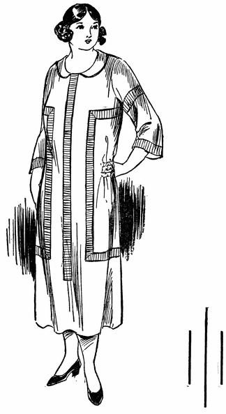
 The middle lines in the two small diagrams are the same length. But on the left, shorter accompanyin...") Optical Illusion in dress
Optical Illusion in dress
The middle lines in the two small diagrams are the same length. But on the left, shorter accompanying lines seem to shorten the one between. On the right longer accompanying lines seem to lengthen the one between.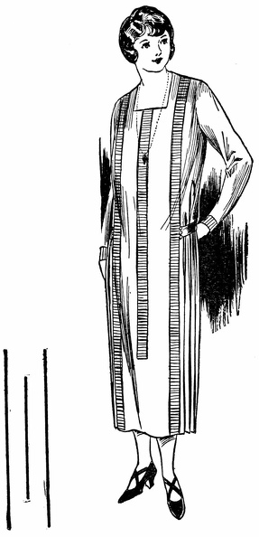
 Now note how these same principles used in the dresses above effect the apparent size and weight of ...") Optical Illusion in dress
Optical Illusion in dress
Now note how these same principles used in the dresses above effect the apparent size and weight of those wearing them, making one seem much stouter than the other.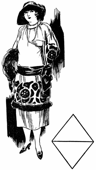
 These two diamond-shaped figures are exactly the same size. The crosswise line makes one seem wider,...") Optical Illusion in dress
Optical Illusion in dress
These two diamond-shaped figures are exactly the same size. The crosswise line makes one seem wider, the vertical line makes the other seem narrower.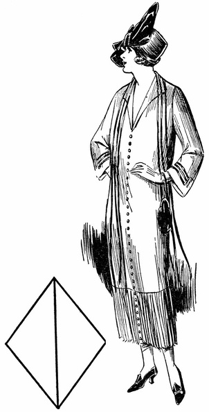
 Here, also, are two vertical parallel lines. They are straight—test them—but the other lines rad...") Optical Illusion in dress
Optical Illusion in dress
Here, also, are two vertical parallel lines. They are straight—test them—but the other lines radiating from the center, make them appear “bowed.” In the dress above a similar design makes the wearer appear stouter and heavier than she really is.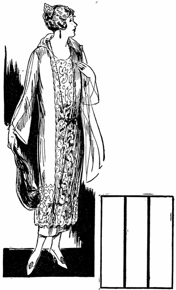
 These unbroken parallel vertical lines give the definite impression of height. This principle, used ...") Optical Illusion in dress
Optical Illusion in dress
These unbroken parallel vertical lines give the definite impression of height. This principle, used in the design of the dress above, lends it a pleasing slender appearance because no other lines interfere with the straight line effect.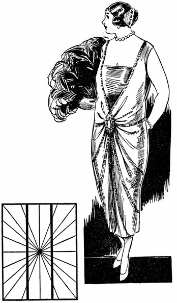
 These two illusions are almost duplicated in the dresses above. As a result one woman looks shorter ...") Optical Illusion in dress
Optical Illusion in dress
These two illusions are almost duplicated in the dresses above. As a result one woman looks shorter and heavier, the other taller and slenderer than she really is.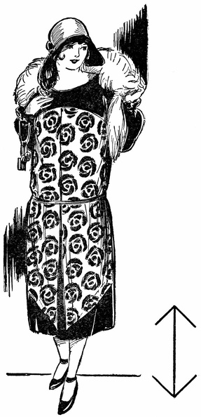
 The two vertical lines are exactly the same length—measure them and see. Short lines turned back a...") Optical Illusion in dress
Optical Illusion in dress
The two vertical lines are exactly the same length—measure them and see. Short lines turned back at either end make one seem short; extended lines make the other seem longer.