


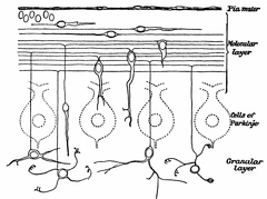
 The Growth and Migration of Granules of the Cerebellum
 The Body of a Motor Neurone
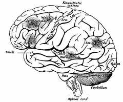 The Surface of the Left Cerebral Hemisphere, Cerebellum,and Medulla Oblongata.
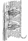 Highly Magnified Section through the Wall of a Circumvallate Papilla of the Tongue, showing Two Taste-Bulbs.
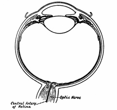 Horizontal Section through the Right Eye
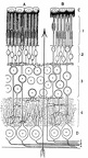 The Retina in Vertical Section
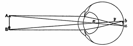 The Formation of an Image by the Refracting Media of the Eye
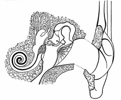 The External, Middle, and Internal Ear of the Left Side
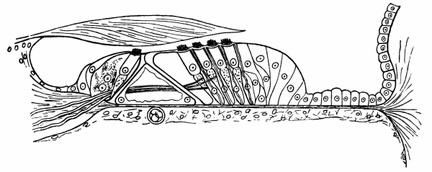 Organ of Corti
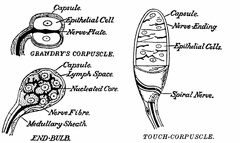 Sense-Organs susceptible to Pressure
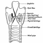 The Anterior Half of the Larynx seen from Behind
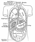 Diagram showing the Relative Positions of the Organs of the Chest and Abdomen.
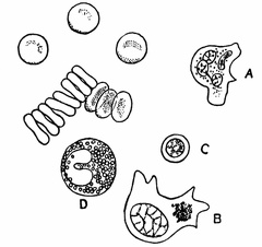 Red Blood-Corpuscles presenting, some the Surfaces, others the Edges, of their Discs, together with Single Representatives of Four Types of Leucocyte.
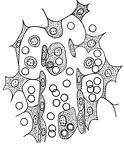 A Minute Portion of the Pulp of the Spleen
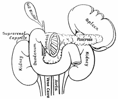 Digestive system in humans
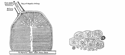 Diagram of a Lobule of the Liver
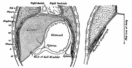 The Diaphragm and Organs in Contact with it
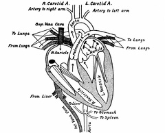 The Heart cut in the Plane of its Long Axis, and the Vessels which open into and out of it
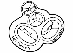 A Section approximately at Right Angles to the Long Axis of the Heart
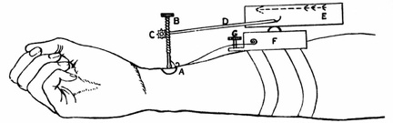 Sphygmograph
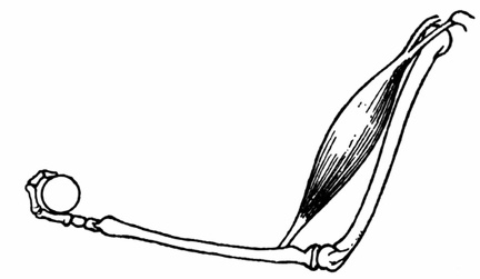 Biceps Muscle in Action