


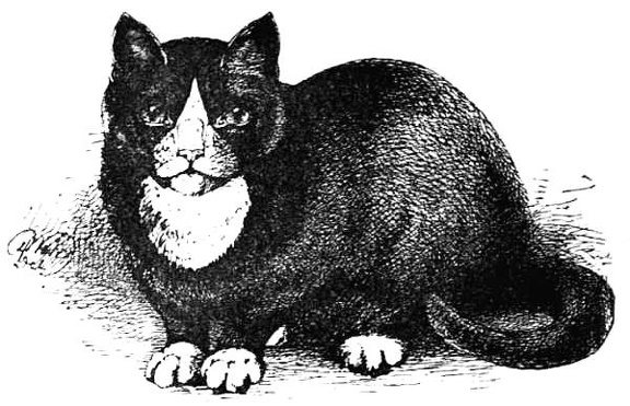
 Preperly Marked Black and White
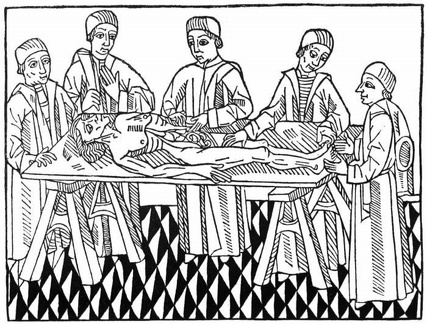 The first printed picture of dissection
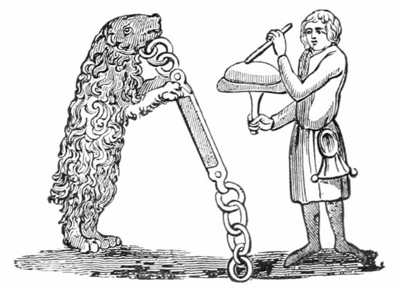 Tutored Bear.—XIV. Century
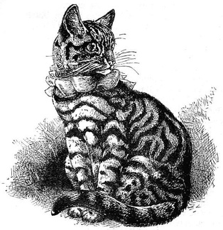 Well-marked Silver Black-banded Tabby
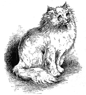 'The Colonel' - White Persian
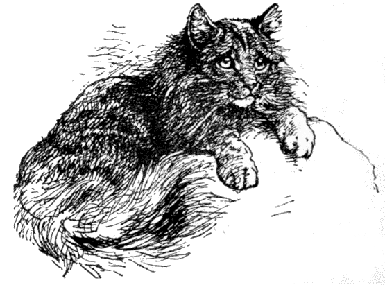 'Tiger'
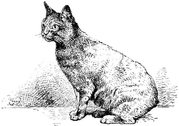 Tortoise Shell Manx
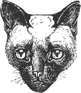 Siamese winner of many prizes
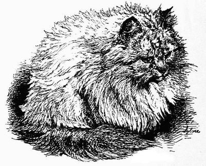 very Light Blue Tabby, 'Sylvie'.
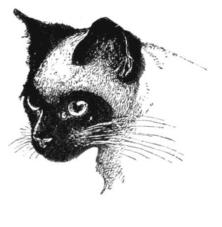 Properly Marked Siamese
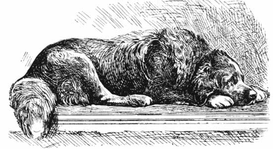 Dog Sleeping
 Lion
Lion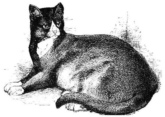 Properly marked black and white cat
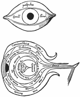 The Anatomy of the Eye
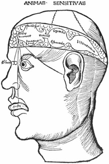 Diagram of the ventricles and the senses
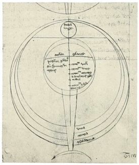 Roger Bacons diagram of the Eye
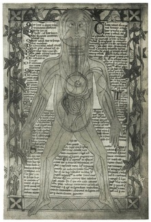 An anatomical diagram of about 1298
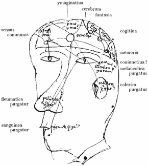 Diagram of the senses, the humours, the cerebral ventricles, and the intellectual facultie
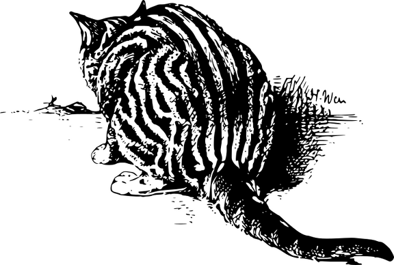 'The old Lady' - Silver Tabby
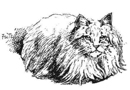 'Fez' - Persian
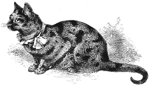 Example of a finely-marked Tortoiseshell Cat
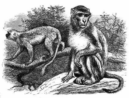 The Rhesus and Entellus. (1 Kings 10. 22)
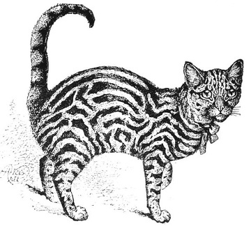 Example of a properly-marked Brown Tabby
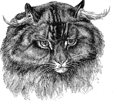 Unusual Long Haired Cat
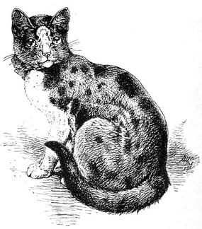 Tortoiseshell-and-white Cat, finely marked
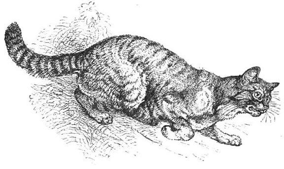 English Wild Cat
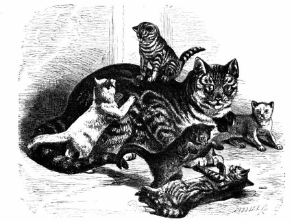 Cat with kittens
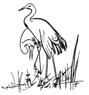 The Two White Birds
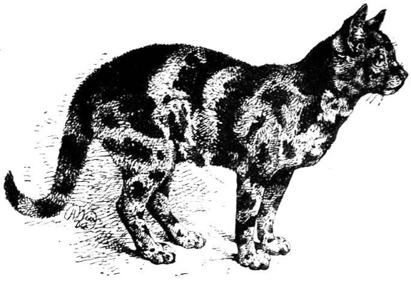 Example of Tortoiseshell Cat, very dark variety
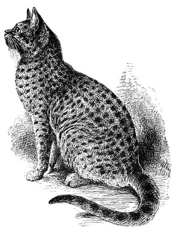 Example of a finely-marked Spotted Tabby He-Cat
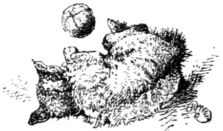 Game of Ball
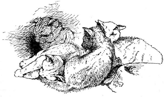 Group of Kittens at the Crystal Palace Cat Show
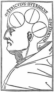 Venice, 1496, showing the ventricles of the brain
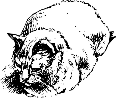 Cat at Show
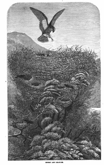 Osprey and Grakles
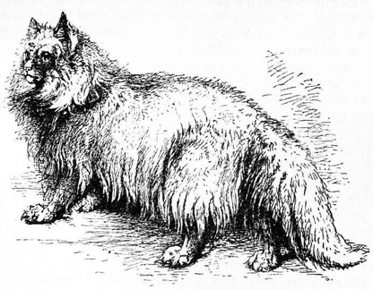 White Angora
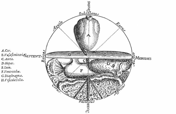 The Microcosm
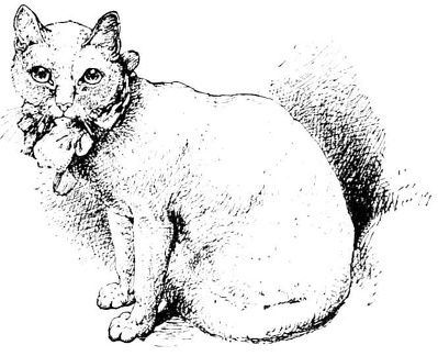 White cat - prize winner in 1879
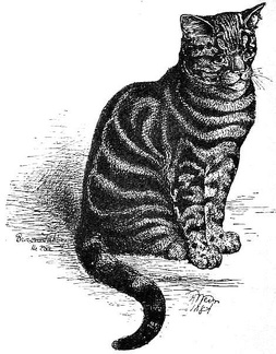 Brown Tabby with the black bars far too wide
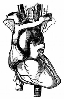 The arch of the aorta and its branches
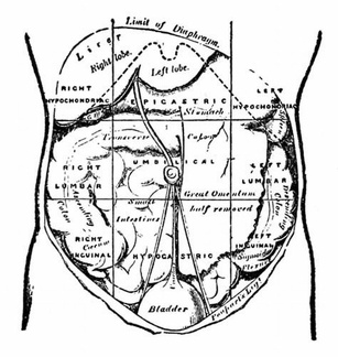 The regions of the abdomen and their contents
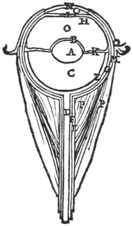 The Anatomy of the Eye
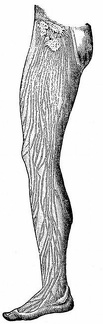 Lymphatics of the leg.
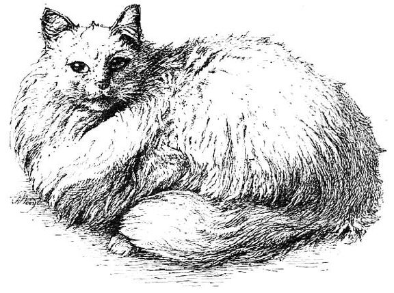 White Persian 'Miss Whitey'
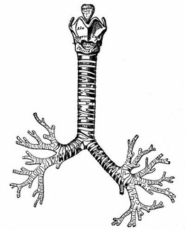 The cartilages of the larynx; the trachea and bronchi
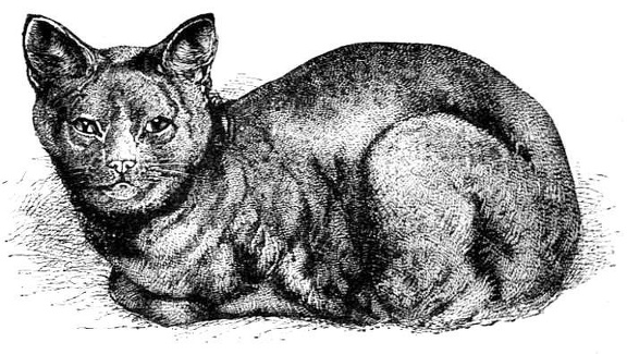 Archangel Blue Cat
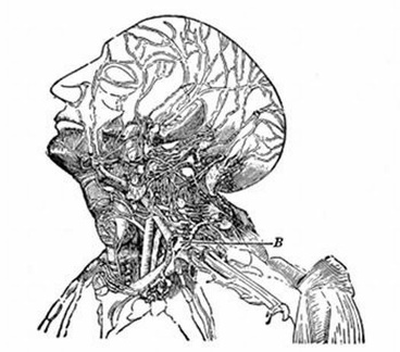 Lymphatics of the head and neck. B, the thoracic duct
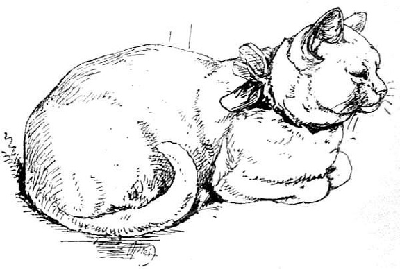 White Cat, winner of many prizes
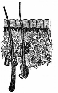 A cross section of the skin
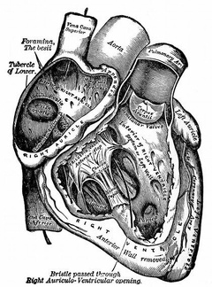 The right auricle and ventricle laid open
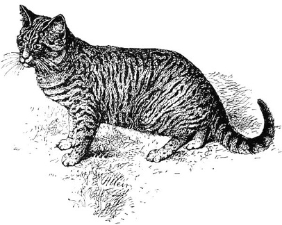 Dark Blue, Small-banded Tabby
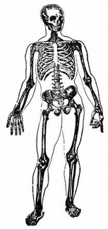 Skeleton
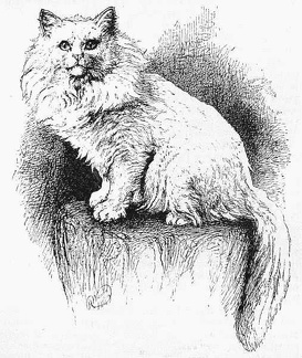 White Persian 'Tim'
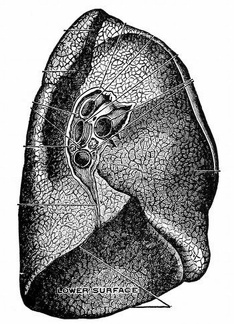 The root of the left lung
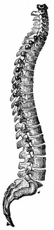 The Spine
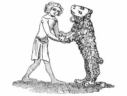 Tutored Bear.—XIV. Century
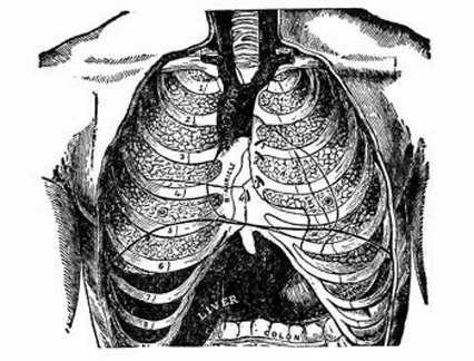 Front view of the thorax
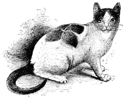 Curiously marked white and black cat
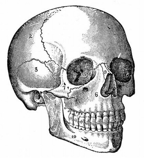 The Skull
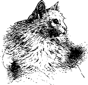 a white Persian - Muff