


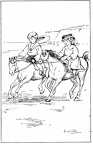
 Two children riding ponies on the beach
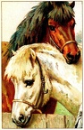 Two horses
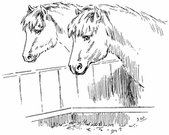 Two horses looking at their food
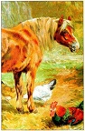 Horse and chickens
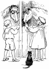 Boy and girl feeding the horses
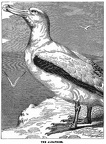 The Albatross
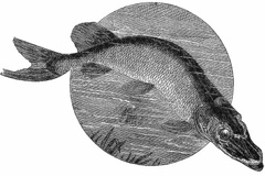 Pike
Pike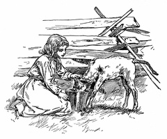 Feeding a goat
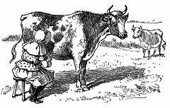 Milking a cow
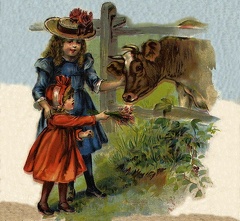 Two girls feeding a cow
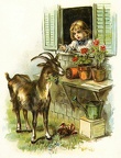 Naughty goat!!
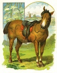 A horse
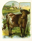 A Cow
A Cow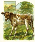 A calf
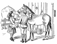 Boy and Girl feeding a horse
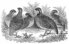 Partridges
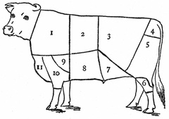 Cow Parts
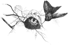 A Clever Humming-bird
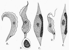 Trypanosoma Ziemanni, from the blood of the little owl
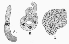 Trypanosoma Ziemanni, from the gut of the gnat
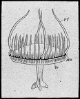 The Freshwater Jelly-fish of Regent’s Park (Limnocodium Sowerbii)
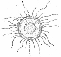 The Freshwater Jelly-fish of Lake Tanganyika
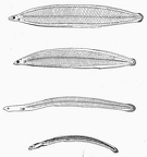 The young of the common Eel and its metamorphosis
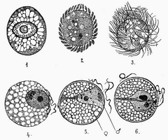 The unicellular parasite Benedenia, from the gut of the common Poulp or Octopus
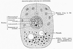 Diagrammatic representation of the structures present in a typical cell
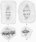 The Number of the Chromosomes
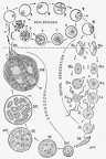 A diagram showing the life-history and migration of the Malaria parasite
 Lankesterella ranarum (Lank.), the parasite of the red blood-corpuscles of the edible Frog
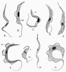 Various species of Trypanosoma from the blood of mammals, birds, and reptiles
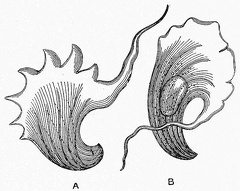 The earliest discovered Trypanosome, described by Gruby in 1843
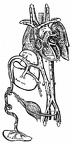 Plan of the foetal circulation
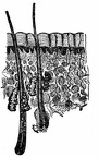 A cross section of the skin
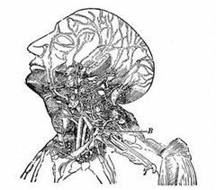 Lymphatics of the head and neck. B, the thoracic duct
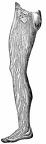 Lymphatics of the leg.
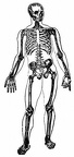 Skeleton
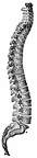 The Spine
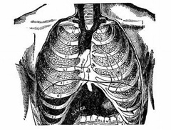 Front view of the thorax
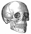 The Skull
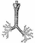 The cartilages of the larynx; the trachea and bronchi
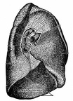 The root of the left lung
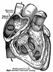 The right auricle and ventricle laid open
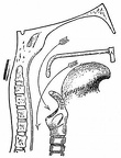 Passage into trachea and esophagus; Pharynx
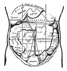 The regions of the abdomen and their contents
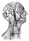 Superficial veins of the head and neck
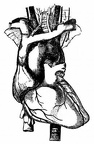 The arch of the aorta and its branches
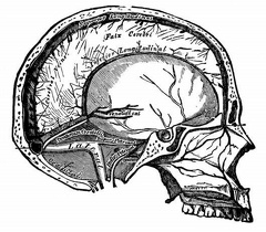 Vertical section of the skull, showing the sinuses of the dura mater
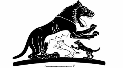 Lioness and young, from an Ionian vase of the sixth century B. C
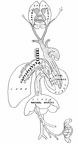 Illustrating Galen’s physiological teaching
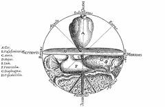 The Microcosm
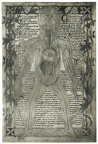 An anatomical diagram of about 1298
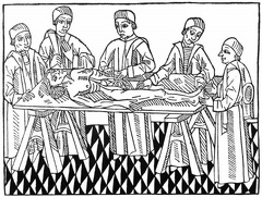 The first printed picture of dissection
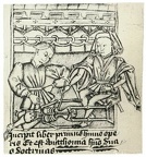 The figure shows a professor and pupil. The former is demonstrating the bones of a skeleton.
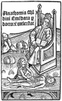 Title-page of Mellerstadt’s edition of the Anatomy of Mondino, Leipzig, 1493. The scene is laid in the open air
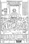 A dissection scene
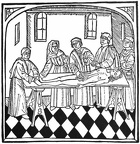 The first picture of dissection in an English-printed book
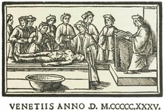 a lecture on anatomy
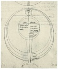 Roger Bacons diagram of the Eye
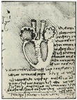 Leonardo Da Vincis diagram of the heart
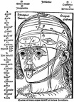 The figure shows the ten layers of the head
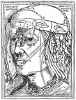 The layers of the head